

CLINICAL SUPPORT
Expert Clinical Histology Services,
designed to deliver precision and reliability
Introduction
At Splice Histology, we offer a complete range of histology services designed to deliver precision and reliability at every step of the workflow. From initial specimen handling to advanced imaging and analysis, we ensure accuracy and efficiency in processing biological tissues for diagnostic and research needs.


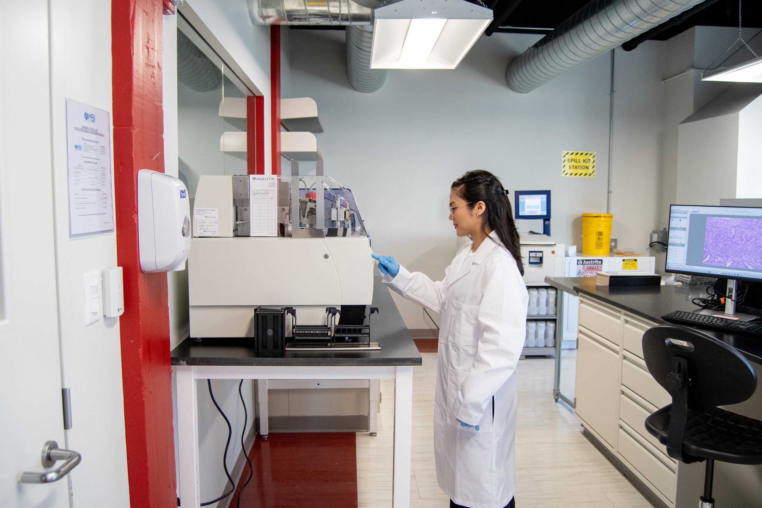
PROCESSING
Description:
Tissues are dehydrated, cleared, and infiltrated with paraffin to preserve structure and prepare for sectioning.
Workflow Details:
1. Tissues undergo dehydration in graded alcohol solutions.
2. Clearing agents are applied to replace alcohol and improve paraffin penetration.
3. Paraffin wax is infiltrated under vacuum to ensure complete saturation.
Duration: 2–12 hours, depending on specimen size and type.
Purpose:
To solidify the tissue for cutting while maintaining cellular integrity.
EMBEDDING
Description:
Tissues are encased in paraffin to create a stable block for sectioning.
Workflow Details:
1. Molten paraffin is poured into embedding molds containing tissue samples.
2. Specimens are oriented to preserve diagnostic areas for sectioning..
3. Blocks are cooled and solidified for stability.
Purpose:
To ensure precise slicing and maintain tissue architecture.
SECTIONING
Description:
Thin slices of tissue are cut using a microtome and placed on glass slides.
Workflow Details:
1. Paraffin blocks are mounted onto a microtome.
2. Tissues are sliced into 3–5 µm sections.
3. Sections are floated on a water bath to ensure smooth placement.
4. Glass slides are used to collect tissue sections.
Purpose:
To create uniform tissue layers suitable for microscopic analysis.
STAINING
Description:
Tissue sections are stained to highlight cellular structures for examination.
Workflow Details:
1. Hematoxylin is applied to stain nuclei purple.
2. Eosin is used to color cytoplasm and extracellular matrix pink.
3. Optional special stains or immunohistochemical (IHC) protocols can be added for specific diagnostic purposes.
Purpose:
To enhance contrast and make tissue morphology discernible.
-
Hematoxylin and Eosin (H&E) staining is a cornerstone in histology, offering a clear view of tissue structure and cellular organization. Hematoxylin binds acidic components like DNA, staining nuclei blue-purple, while eosin highlights proteins in the cytoplasm and extracellular matrix in shades of pink. This contrast enables pathologists to assess tissue morphology and identify conditions like inflammation, necrosis, or malignancy. Its simplicity and reliability make it indispensable for diagnostic applications.
-
Immunohistochemistry (IHC) detects specific proteins in tissues using antigen-antibody binding. A primary antibody targets the antigen, while an enzyme-linked secondary antibody generates a visible color reaction. IHC is crucial for diagnosing diseases and characterizing markers like HER2 in breast cancer. Combining specificity with semi-quantitative analysis, it provides critical insights into protein expression patterns in clinical and research settings.
-
Immunofluorescence (IF) staining uses fluorophore-conjugated antibodies to detect proteins in tissues or cells. Fluorophores emit light when excited, enabling precise localization of proteins under a fluorescence microscope. IF allows detailed study of protein expression and interactions, often using multiple markers simultaneously. Known for its sensitivity and spatial resolution, it is widely used in research, although careful optimization is needed to avoid signal overlap or fading.
-
Multiplex staining enables detection of multiple targets in a single tissue sample. Brightfield multiplexing uses chromogenic dyes visible under a light microscope, ideal for routine diagnostics. Fluorescent multiplexing employs fluorophores with distinct emissions, allowing high-sensitivity analysis of complex cellular environments, such as the tumor microenvironment. Techniques like spectral unmixing enhance its capacity for highly multiplexed and detailed investigations in research.
-
Histochemical staining identifies biochemical properties of tissues through direct chemical reactions. Examples include PAS for carbohydrates, Alcian Blue for mucins, and Prussian Blue for iron. These stains are essential for diagnosing metabolic and storage disorders, as well as identifying tumor features. Cost-effective and simple, histochemical methods complement structural and biochemical tissue analysis effectively.
OUR STAINING CATEOGORIES
COVERSLIPPING
Description:
Glass coverslips are applied to slides to protect tissue sections and prepare them for microscopy.
Workflow Details:
1. Mounting media is applied to slides for adhesion.
2. Coverslips are placed to minimize air bubbles and ensure longevity.
3. Slides are labeled and archived for analysis.
Purpose:
To protect samples and provide optimal viewing conditions under a microscope.
SLIDE IMAGING
Description:
Tissue sections are digitized for advanced analysis and remote consultation.
Workflow Details:
1. Slides are scanned using high-resolution imaging systems.
2. Digital images are processed and stored for review.
3. Remote access is enabled for pathologists and researchers.
Purpose:
To modernize histology workflows and facilitate collaboration.
KEY FEATURES
Description:
Tissue specimens are visually inspected and measured. Key areas of interest are identified and sectioned for further processing.
Quality Assurance:
Each stage undergoes strict quality checks to maintain precision.
Customization:
Services can be tailored to meet client-specific needs.
24/7 Availability:
Our lab operates around the clock to meet urgent demands.